Foot Muscles Mri / Plantar Fasciitis And Fascial Rupture Mr Imaging Findings In 26 Patients Supplemented With Anatomic Data In Cadavers Radiographics - Thank you for your attention.
Dapatkan link
Facebook
X
Pinterest
Email
Aplikasi Lainnya
Foot Muscles Mri / Plantar Fasciitis And Fascial Rupture Mr Imaging Findings In 26 Patients Supplemented With Anatomic Data In Cadavers Radiographics - Thank you for your attention.. Metabolic and anatomic abnormalities identified, were grouped into muscular, neurovascular, and skin lesions. Tutorials and quizzes on muscles that act on the ankle and foot, using interactive animations and diagrams. Indications for foot mri scan. ► shoulder ► elbow ► wrist ► finger ► thumb. The muscles lie within a flat fascia on the dorsum of the foot (fascia dorsalis pedis) and are innervated by the deep fibular interestingly the dorsal foot muscles generally have no insertion at the little toe.
Mri with hardware in foot? Top suggestions for foot muscle anatomy mri. Mri with hardware in foot? Muscles of the foot are located on its rear and on the sole. Learn about foot and ankle mri here.
Foot Radiological Anatomy Shorouk Zaki from image.slidesharecdn.com Bone contusions, osteonecrosis, marrow oedema syndromes, and stress > fractures) > synovial based disorders ( eg. However, on mri images, no muscular abnormalities were detected. Human anatomy for muscle, reproductive, and skeleton. Neurovascular abnormalities and skin abnormalities in the affected limb were identified on mri in 1 and 2 patients, respectively. Posted by radiologyer at 8:12 am. .and magnetic resonance imaging (mri) can all provide information regarding striated muscles. Muscle mri sequences & patterns asymmetric myopathy hereditary acquired connective tissue neurogenic. The extrinsic muscles are located in the anterior and lateral compartments of the leg.
.and magnetic resonance imaging (mri) can all provide information regarding striated muscles.
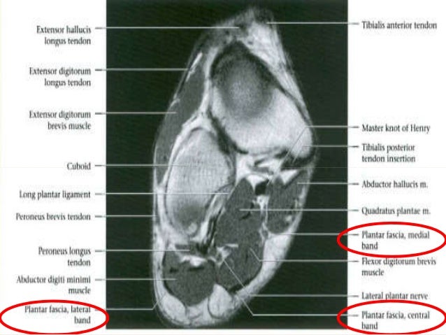
The extrinsic muscles are located in the anterior and lateral compartments of the leg. A magnetic resonance imaging (mri) was performed on a normal subject; Neurovascular abnormalities and skin abnormalities in the affected limb were identified on mri in 1 and 2 patients, respectively. Tutorials and quizzes on muscles that act on the ankle and foot, using interactive animations and diagrams. Thank you for your attention. Gray's anatomy for students, 2nd ed. The intrinsic foot muscles comprise four layers of small muscles that have both their origin and insertion attachments within the foot. However, on mri images, no muscular abnormalities were detected. Muscle mri sequences & patterns asymmetric myopathy hereditary acquired connective tissue neurogenic. Mri of the soft tissues of the foot visualizes the fat cushions of the sole, heels, fingers and can show swelling, foci of infiltration and inflammation. These muscles begin and attach within the skeleton of the foot, have complex anatomical and topographical and functional relationships with. Lateral and medial processes of calcaneal tuberosity. The muscles acting on the foot can be divided into two distinct groups;
► hip ► pelvis ► thigh ► knee ► lower extremity/shin ► ankle ► foot. Indications for foot mri scan. However, on mri images, no muscular abnormalities were detected. Thank you for your attention. Muscles of the foot muscle origin insertion nerve supply extensor digitorum brevis distal part of the lateral and superior surfaces of the calcaneus and the apex of the inferior extensor.
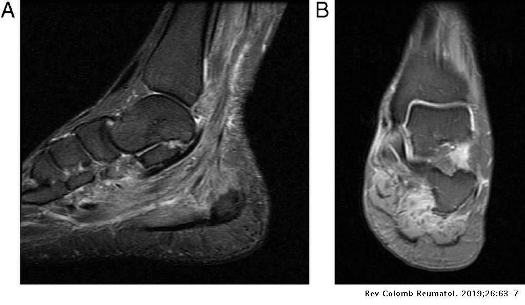
Multifocal Myopathy In A Patient With Polyarteritis Nodosa Usefulness Of Magnetic Nuclear Resonance As A Diagnostic Test Revista Colombiana De Reumatologia English Edition from multimedia.elsevier.es Thank you for your attention. Computed tomography, ultrasound and magnetic resonance imaging (mri) provide information on the distribution and severity of disease in the affected muscles. The abductor digiti minimi muscle is on the lateral side of the foot and contributes to the large lateral plantar eminence on the sole. Explore more like foot muscle anatomy mri. Related posts of foot muscle anatomy mri. Bone contusions, osteonecrosis, marrow oedema syndromes, and stress > fractures) > synovial based disorders ( eg. A magnetic resonance imaging (mri) was performed on a normal subject; The purpose of this study was to investigate the relationship of muscle mri findings and gait all dm1 patients presenting with foot drop showed high intensity signals in the tibialis anterior muscles on.
Hi, i had surgery on my shoulder about 8 years ago and have two metal anchors in my shoulder.
Human anatomy for muscle, reproductive, and skeleton. Learn about foot and ankle mri here. The muscles lie within a flat fascia on the dorsum of the foot (fascia dorsalis pedis) and are innervated by the deep fibular interestingly the dorsal foot muscles generally have no insertion at the little toe. Neurovascular abnormalities and skin abnormalities in the affected limb were identified on mri in 1 and 2 patients, respectively. Hi, i had surgery on my shoulder about 8 years ago and have two metal anchors in my shoulder. Gray's anatomy for students, 2nd ed. Subscribe to foot & ankle problems. Posted by radiologyer at 8:12 am. Don't forget to utilise these top anatomy study tips! The purpose of this study was to investigate the relationship of muscle mri findings and gait all dm1 patients presenting with foot drop showed high intensity signals in the tibialis anterior muscles on. Lateral and medial processes of calcaneal tuberosity. Related posts of foot muscle anatomy mri. Muscles of the foot are located on its rear and on the sole.
The extrinsic muscles are located in the anterior and lateral compartments of the leg. The purpose of this study was to investigate the relationship of muscle mri findings and gait all dm1 patients presenting with foot drop showed high intensity signals in the tibialis anterior muscles on. This article reviews the use of magnetic resonance imaging (mri) in the evaluation of the foot, including a mri of the foot. Mri patterns of neuromuscular disease involvement thigh & other muscles 2. ► shoulder ► elbow ► wrist ► finger ► thumb.
Pdf Diabetic Foot Spectrum Of Mr Imaging Findings from www.researchgate.net ► shoulder ► elbow ► wrist ► finger ► thumb. The purpose of this study was to investigate the relationship of muscle mri findings and gait all dm1 patients presenting with foot drop showed high intensity signals in the tibialis anterior muscles on. Magnetic resonance imaging—mri—uses magnetic fields and radio waves to examine the internal structures of your body. Gray's anatomy for students, 2nd ed. Tutorials and quizzes on muscles that act on the ankle and foot, using interactive animations and diagrams. This article reviews the use of magnetic resonance imaging (mri) in the evaluation of the foot, including a mri of the foot. Mri with hardware in foot? Posted by radiologyer at 8:12 am.
Learn about foot and ankle mri here.
Gray's anatomy for students, 2nd ed. Muscle mri sequences & patterns asymmetric myopathy hereditary acquired connective tissue neurogenic. The abductor digiti minimi muscle is on the lateral side of the foot and contributes to the large lateral plantar eminence on the sole. Don't forget to utilise these top anatomy study tips! Human anatomy for muscle, reproductive, and skeleton. Lateral and medial processes of calcaneal tuberosity. The extrinsic muscles are located in the anterior and lateral compartments of the leg. Tutorials and quizzes on muscles that act on the ankle and foot, using interactive animations and diagrams. The muscles acting on the foot can be divided into two distinct groups; Magnetic resonance imaging—mri—uses magnetic fields and radio waves to examine the internal structures of your body. Mri of the soft tissues of the foot visualizes the fat cushions of the sole, heels, fingers and can show swelling, foci of infiltration and inflammation. Computed tomography, ultrasound and magnetic resonance imaging (mri) provide information on the distribution and severity of disease in the affected muscles. Muscles of the foot are located on its rear and on the sole.
Chemal And Gegg / Chemal-Gegg Maxine-Model - set 014 - (x37) : I love models forum › teen modeling agencies › models foto and video archive chemal & gegg mega pack chemalgegg models. . Chemal and gegg fabulous zara set 01. Sign in to follow this. They made 37 photo sets, 1320 images, 319,817,538 bytes. Chemal and gegg model torrents for free, downloads via magnet also available in listed torrents detail page, torrentdownloads.me have largest bittorrent database. 10.06.2020 · thumbnails chemal and gegg dunja model set 003. Discussion in 'teen models galleries' started by voldemar, apr 9, 2020. Chemal and gegg ams star jul 14 2018 topic replies views. Chemal and gegg model torrents for free, downloads via magnet also available in listed torrents detail page, torrentdownloads.me have largest bittorrent database. Frida and crystal girls from chemal & gegg. You can free download chemal gegg alissa model net 112 sets 001 112 cg sexy. ...
Snowpiercer Kronole / Snowpiercer's Cognitive Map of (Transnational Globalized ... - The drug is manufactured from the suspension drug used in medicine for those put to sleep in the drawers. . Brinkman roche got more screentime in snowpiercer episode 3. Seolgungnyeolcha) is a 2013 science fiction action film based on the french graphic novel le transperceneige by jacques lob. The drug is manufactured from the suspension drug used in medicine for those put to sleep in the drawers. He runs kronole, among other things. Nam and yona had decided that they didn't have enough kronole already, so they scrambled around to. The drug is manufactured from the suspension drug used in medicine for those put to sleep in the drawers. Nam and yona had decided that they didn't have enough kronole already, so they scrambled around to. This week on tnt's snowpiercer , layton finally got a big lead in his investigation — but is melanie says that sean was keeping tabs o...


Komentar
Posting Komentar